Chromosome
n., plural: chromosomes
[ˈkrəʊməˌsəʊm]
Definition: a cell structure that bears the genetic material
Table of Contents
Chromosomes Definition
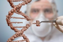
Chromosomes are thread-like structures present in the nucleus of plant and animal cells. Chromosomes are the vehicle that carries DNA (the genetic material of the cell) wrapped around by the histone proteins.
To easily understand what a chromosome is, let us go through this list of the most commonly asked questions about chromosomes.
- Question: What are chromosomes and DNA? Chromosomes are threadlike structure that carries DNA along with histone protein. Chromosomes are composed of DNA, and the primary chromosome function is to carry the essential genetic material of the cell, i.e., DNA holds all the genetic information encoded in DNA genes. Does this also clarify what are genes made of? Genes are basically tiny part of DNA that stores genetic information encoded in nucleotide sequences.
Question: Where are chromosomes located? Or where are chromosomes found? Well, the location of chromosomes depends upon the type of the cell. For example, the eukaryotic chromosome is present in the nucleus, while in prokaryotes, chromosomes are present in the cytoplasm in the nucleoid region of the cell. Chromosomes are present in the nucleus and not in the cell’s nucleolus.
Question: Where is the nucleolus found? Well, the nucleolus is the cell organelle present in the nucleus that carries out the function of producing and gathering ribosomes.
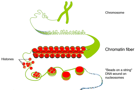
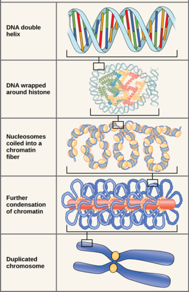
Question: What is a chromosome? What is it made up of? Are chromosomes made of DNA? The answer to it is yes, chromosomes are made up of DNA. Essentially, chromosomes are made up of DNA packed around histone proteins forming a structure known as nucleosomes. Further, these nucleosomes coil up tightly to form chromatin loops. These chromatin loops further coil up to form chromosomes. The methylation of DNA and other intermolecular forces helps to keep the chromosomes in a stable coiled 3-D tertiary structure. The tertiary structure of the chromosomes is critical for cell division and regulation of transcription and cellular replication.
Question: Where is DNA found in a cell? It depends, DNA can be found in the nucleus of the eukaryotic cell. It may also be found in other organelles, such as mitochondria and chloroplasts. In a prokaryotic cell, it is found in the nucleoid region of the cytoplasm.
Question: What are histones? Histones proteins are rich in lysine and arginine residues and possess a positive charge on them, while DNA has a negative charge on it. Thus, electrostatic interaction between histone protein and DNA aids in the wrapping of the DNA with the histone protein.
Question: How about bacteria and other prokaryotes? What makes their chromosomes different? Distinctly, prokaryotic chromosomes do not possess histone proteins, while eukaryotic chromosomes contain histone proteins.
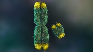
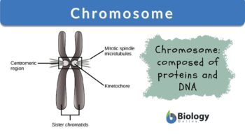
Structurally, each chromosome comprises two long arms (q arm) and two short arms (p arm), i.e., 4 chromosome arms held together at the centromere, resulting in the formation of an ‘X’ shaped structure. However, the position of the centromere determines the chromosomal shape. Accordingly, there are different types of chromosomes, namely,
- Telocentric chromosomes: Herein, the centromere is present at the terminal end of the arm of the chromosome; as a result, there is a shorter arm, and the other is a long arm.
- Acrocentric chromosome: Herein, the centromere is placed near to chromosomal arm end, making the p arm very short while the q arm is very long.
- Submetacentric chromosomes: Wherein the centromere is present almost at the center, but not at the exact center of the chromosomes resulting in the p arm being a little shorter than the q arm.
- Metacentric chromosome: Herein, the centromere is present exactly in the middle of the chromosome, resulting in an equal p and q arm length.
Almost all the body cells of the body possess chromosomes. Chromosomes are pretty long structures spanning nearly 6ft from end to end. However, chromosomes can be visualized under a microscope only in the metaphase stage of cell division when all the genetic material is in a highly condensed state wherein it can be visualized as an X-shaped structure with two sister chromatids attached to each other at a centromeric point or the centromere.
Watch this video to recognize the parts of the chromosome and the types of chromosomes based on the position of the centromere.
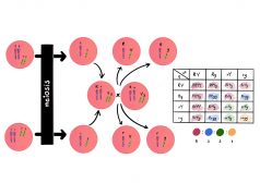
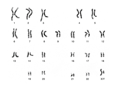
Question: How many chromosomes do humans have? Or What are the 46 chromosomes? The shape and number of chromosomes differ in each living organism. So, in a human cell, there are 23 pairs of chromosomes, i.e. 46 chromosomes wherein half of the chromosomes are from the mother, and the other half is from the father.
Somatic human cells have 23 pairs of chromosomes, i.e., 46 chromosomes. Of these, 22 chromosomes are autosomal, and the 23rd chromosome is the sex chromosome. Autosomal chromosomes are homologous that contain genes encoding similar traits. Such somatic cells are called diploid cells as their nucleus has a pair of homologous chromosomes.
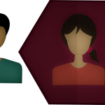
Contrary to this, haploid cells do not possess pair of these chromosomes and contain only one set of chromosomes for, e.g., germ cells, i.e., sperm and egg. Accordingly, diploid cells are represented as 2n, while haploid cells are represented as n. Furthermore, germ cells have either X chromosomes or Y chromosomes. The Y chromosome is relatively smaller in size than the X chromosome and is responsible for imparting male characters, i.e., the Y chromosome is a male chromosome. Therefore, the presence of the XX chromosome results in female offspring while the XY chromosome gender would be male.
Watch this video to learn about human chromosomes and karyotypes.
A chromosome is a structure within the cell that bears the genetic material as a threadlike linear strand of DNA bonded to various proteins in the nucleus of eukaryotic cells, or as a circular strand of DNA (or RNA in some viruses) in the cytoplasm of prokaryotes and in the mitochondrion and chloroplast of certain eukaryotes. Etymology: Greek chroma (“color”) + soma (“body”).
Some chromosomes are homologous; others are nonhomologous. Find out how to tell them apart. Join our Forum: Difference Between Homologous Chromosomes and Sister Chromatids. Discover more!
In eukaryotes, the chromosomes appear as threadlike strands that condense into thicker structures and align on the metaphase plate during mitosis. Humans normally have 23 pairs of chromosomes, each with a characteristic length and banding pattern. Chromosomes occur in pairs (in most somatic cells) since one member of each pair comes from the mother and the other from the father. In most prokaryotes, the chromosome is usually a circular strand of DNA; hence, the entire genome is carried on only one chromosome. In viruses, the chromosome may appear as a short linear or circular structure containing the DNA or RNA molecule often lacking any structural proteins.
History of Discovery
Walther Flemming discovered chromosomes in 1878. He termed the thread-like structure found in the nucleus as ‘chromatin’. While the term ‘Chromosomes’ was coined in 1888 by W. Waldeyer. Reger Kornberg, in 1974 reported that chromosome is made up of DNA and protein.
Prokaryotes
Prokaryotes have a one, coiled, circular (mostly) chromosome present in the nucleoid region of the prokaryotes (as prokaryotes are devoid of a nucleus) [though certain prokaryotes like Vibrio cholerae have two circular chromosomes]. The prokaryotic chromosome lacks histone protein, but it is supercoiled to form a loop. The supercoiling of the chromosomes in prokaryotes is aided by nucleoid-associated proteins (NAPs). Therefore, prokaryotic cells are haploid cells, i.e., they possess only one chromosome.
Even prokaryotes like Vibrio cholerae are haploid as the two circular chromosomes present in them are not homologous. Hence, Vibrio cholerae is a haploid cell. Apart from the chromosome, some prokaryotes have additional or extra-chromosomal DNA, which is known as a plasmid. Plasmids are an important biotechnological tool used to synthesize bacterial artificial chromosomes.
Watch this video to learn the difference between prokaryotic and eukaryotic chromosomes
Structure in sequences
A prokaryotic chromosome is devoid of introns and has highly conserved co-regulated genes. These clusters of co-regulated genes are known as operons. Though their genetic sequence may differ, these co-regulated genes operate for the same function. Usually, the genes in the same functional pathway are coded by operons. For example, lac operon in E. coli– lacZ, lacY, and lacA – all three contribute to lactose utilization in E.coli. Such a coregulated gene cluster helps bacteria adapt to environmental changes better and rapidly.
DNA packaging
Chromosomes are pretty long in prokaryotes; for example, almost 4.6 million base pairs, spanning a length of approximately 1.1mm, are found in E.coli. It is pretty intriguing how such a long structure is packed into a small cell. DNA gets coiled and twisted to form a supercoiled ball-like structure for this to happen. To better understand it, one can imagine a twisted rubber band to form a small coil-like structure; upon further coiling such a rubber band, it forms a supercoiled ball-like structure. Similarly, the prokaryotic DNA is coiled in the chromosome. Now, this coiling of DNA can occur as:
- Under wound, i.e., less than one helical turn/10 base pair
- Over wound- more than one helical turn/10 base pair
Further, this coiling of DNA can occur in the same direction as the DNA double helix (i.e., positively supercoiled), or it can occur in the opposite direction to that of the DNA helix (i.e., negatively supercoiled). The majority of the bacterial genome is negatively supercoiled during the normal growth phase.
One may wonder how transcription occurs in such a supercoiled structure. It has been observed that small projections appear from the nucleoid during the transcription process. These projections seem to allow easy access to DNA for the transcription process. Interestingly, these projections retract and disappear once the transcription process is finished.
NAPs and enzymes like DNA gyrase and topoisomerase I help in maintaining this supercoiled structure in a prokaryotic cell. Interestingly, Archaeans are the only protozoan group that possesses histone protein-like proteins for condensing their DNA in the chromosome.
Eukaryotes
Eukaryotic DNA is composed of DNA and histone protein. DNA is a long structure that needs to be packed in a small cellular nucleus (human cellular DNA is approximately 2 m in length). Hence, DNA is wrapped around histone proteins. At the primary packaging stage, short DNA stretches are wrapped around the core of 8 histone proteins at regular intervals. These DNA-histone complexes are known as chromatin.
Chromatins appear as bead-like structures on the linker DNA, known as a nucleosome. Nucleosomes further condense and coil to shorten the chromosome by almost 50 times. Finally, it is additionally packed with the help of fibrous protein. However, the extent of DNA coiling and condensation differs according to different cell division stages.
Interphase chromatin
The interphase is the non-dividing stage of the cell. During this phase, the chromatin is less organized and is moderately decondensed as a result, the chromatin is spread throughout the nucleus. At this stage, a part of DNA is decondensed in the transcriptionally active stage and known as euchromatin, while 10% of the chromatin is transcriptionally inactive and is highly condensed, and is known as heterochromatin. Non-condensed DNA state allows the transcription process. Heterochromatin contains a large number of repetitive DNA sequences similar to those found in telomeres and centromeres.
Metaphase chromatin and division
As the cell enters the dividing stage, i.e., metaphase 1, the chromosomes become highly condensed structure. This is to ensure that it will be distributed to the daughter cells. By an estimate, the DNA folds by almost 10,000 fold at the metaphase stage. At this stage, the process of transcription stops. Also, this is the only stage when chromosomes can be visualized under a light microscope.
Human chromosomes
Each species possesses a characteristic number of chromosomes. Humans possess a pair of 23 chromosomes. Of these 23 pairs of chromosomes, 22 pairs of chromosomes are autosomal, i.e., genes present on these chromosomes are not related to sex. These autosomal chromosomes are the same in males as well as females. While the 23 rd chromosome pair is the sex chromosome, i.e., male and female chromosomes. The sex chromosome pair is XY in males, while the sex chromosome pair is XX in females. The X chromosome size is larger than the Y chromosome. The X chromosome has around 2000 genes, while the Y chromosome has around 100 genes. Also, it is essential to note that the Y chromosome determines the sex of the offspring.
Number in Various Organisms
Let’s find out the different numbers of chromosomes of various organisms.
In eukaryotes
Different organisms have different numbers of chromosomes. Some examples of chromosomes in eukaryotic cells are below
Table 1: Number of chromosomes of different eukaryotes |
||
|---|---|---|
| Organism | Diploid number of chromosomes (2n) | Number of chromosomes |
| Humans | 23 | 46 |
| Earthworm | 18 | 36 |
| Dogs | 39 | 78 |
| Lily | 12 | 24 |
| Frog | 13 | 26 |
| Turkey | 41 | 82 |
| Shrimp | 127 | 254 |
| Chimpanzee | 24 | 48 |
| Potato (Solanum tuberosum) |
24 | 48 |
| Hare | 24 | 48 |
| Zebrafish (Danio rerio) |
25 | 50 |
| Water buffalo (Riverine type) (Bubalus bubalis) | 25 | 50 |
| Striped skunk
(Mephitis mephitis) |
25 | 50 |
| Pineapple (Ananas comosus) |
25 | 50 |
| Giraffe (Giraffa camelopardalis) |
15 | 30 |
| Pistachio (Pistacia vera) |
15 | 30 |
| Sunflower (Helianthus annuus) |
17 | 34 |
| Porcupine (Erethizon dorsatum) |
17 | 34 |
| Peanut (Arachis hypogaea) |
20 | 40 |
| American beaver (Castor canadensis) |
20 | 40 |
| Wheat (Triticum aestivum) |
21 | 42 |
| Rhesus monkey (Macaca mulatta) |
21 | 42 |
| Rat (Rattus norvegicus) |
21 | 42 |
| Oats (Avena sativa) |
21 | 42 |
| Giant panda (Ailuropoda melanoleuca) |
21 | 42 |
| Strawberry (Fragaria × ananassa) |
28 | 56 |
| Elephant (Elephantidae) |
28 | 56 |
| Goat (Capra hircus) |
30 | 60 |
| Donkey (Equus asinus) |
31 | 62 |
| Guinea pig (Cavia porcellus) |
32 | 64 |
| Horse (Equus caballus) |
32 | 64 |
| Sloth bear (Melursus ursinus) |
37 | 74 |
| Polar bear (Ursus maritimus) |
37 | 74 |
| Dove (Columbidae) |
39 | 78 |
| Chicken (Gallus gallus domesticus) |
39 | 78 |
| Sugarcane (Saccharum officinarum) |
40 | 80 |
| Crucian carp (Carassius carassius) |
50 | 100 |
| Kamraj (fern) (Helminthostachys zeylanica) |
47 | 94 |
| Rattlesnake fern (Botrypus virginianus) |
92 | 184 |
| Black mulberry (Morus nigra) |
154 | 308 (highest amongst plants) |
In prokaryotes
The majority of prokaryotes have a single chromosome only; however, Vibrio cholerae has two chromosomes.
Karyotype
In simple words, a karyotype is the image of the total number of chromosomes that an organism possesses. This cytogenetic technique is used to identify the abnormalities in the chromosomes in terms of number, size, or shape.
The traits in any organism are due to the genes present in the DNA of that organism. These genes are passed on from the parents to the offspring. Normal human cells possess 46 chromosomes (23 chromosomes from each of the parents); however, in certain offspring, there is a possibility of change in the number, size, or shape of the chromosome resulting in genetic disorder in the offspring. Karyotyping is carried out in pregnant women to identify any genetic disease in the fetus. Also, human cells have 22 pairs of homologous chromosomes and a pair of sex chromosomes. Identification of sex chromosomes can help to reveal the gender of the offspring. Overall, a karyotype test may be required to:
- Checking for any possible genetic disorder in the unborn fetus
- Testing for any congenital disease in a young individual
- Checking for the possibility of genetic disorder is the reason for repeated miscarriages in any women
- Checking for a genetic disorder in a stillborn child for identifying the cause of death
- Rule out the possibility of any congenital disease in an offspring
Some of the possible genetic diseases are summarized in the table below.
Table 2: Genetic disorder and the associated chromosomal number |
|
|---|---|
| Genetic disorder | Number of chromosomes |
| Down syndrome | Trisomic chromosome 21- Down syndrome chromosome |
| Patau syndrome | Trisomic chromosome 13 |
| Edwards syndrome | Trisomic chromosome 18 |
| Turner syndrome | A female offspring with a damaged X chromosome |
| Klinefelter syndrome | A male offspring with an extra chromosome X, i.e., XXY chromosomes instead of a normal XY chromosome. Also known as 47 XXY syndrome. |
| Cri du chat | An example of aneuploid, Trisomy of chromosome 5 resulting in the abnormal larynx, which gives a distinct cry to the offspring |
| Triple X Syndrome | Offspring with XXX chromosomes |
| DiGeorge syndrome or 22q syndrome | Deletion of the long arm of chromosome number 22 |
| Pentasomy X or 49, XXXXX syndrome | Presence of three extra XXX chromosomes |
| Angelman syndrome (AS) and Prader-Willi syndrome (PWS) | These two are Uniparental disomy disorder. A unique condition wherein instead of one copy of chromosome from the parent, two copies of chromosomes are transferred to the offspring |
| Mosaic Turner syndrome | It is different from Turner syndrome. In mosaic turner syndrome, different body cells have different chromosomal constitutions, some cells have X chromosome while other cells don’t have X chromosome. |
History and analysis techniques
The term karyotype was coined by German anatomist Heinrich von Waldeyer in 1888.
Primarily, WBCs are used in this technique. However, cells from bone marrow, amniotic fluid, and placenta can also be used as a sample. In this technique, the chromosomes are isolated, followed by staining and examination under the microscope. The cells are stained with a dye like Gimesa stain after arresting the cell division at the metaphase stage with the help of colchicine. A trained cytogeneticist then arrange the chromosome labeled as 1-23 according to their size from largest to smallest. In humans, chromosome number 1 is the largest, while chromosome number 18 is the smallest. (Figure 3)
Try to answer the quiz below to check what you have learned so far about chromosomes.

Further Reading
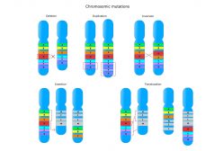
- Chromosome Mutations – Biology Tutorials
- What is Chromatin? Chromosome vs. Chromatin
- Types of Chromosomes: Sex Chromosomes and Autosomes
References
- Stewart F. (2010). The anatomy of a chromosome. The Ulster medical journal, 79(3), 110–113.
- Kuzminov A. (2014). The precarious prokaryotic chromosome. Journal of bacteriology, 196(10), 1793–1806. https://doi.org/10.1128/JB.00022-14
- Osbourn, A. E., & Field, B. (2009). Operons. Cellular and molecular life sciences: CMLS, 66(23), 3755–3775. https://doi.org/10.1007/s00018-009-0114-3
- Fierz B. (2019). Revealing chromatin organization in metaphase chromosomes. The EMBO journal, 38(7), e101699. https://doi.org/10.15252/embj.2019101699
CELL – LABEL THE PARTS (pdf) |
CELL – LABEL THE PARTS
This worksheet is useful in helping the students assess their familiarity with the different parts of the cell, both the eukaryotic and the prokaryotic types. This is useful in genetics as it helps gauge the student’s knowledge of the difference between the two cell types, especially in terms of the location of the genetic material. Subjects: Genetics & Evolution, Cell Biology |
©BiologyOnline.com. Content provided and moderated by BiologyOnline Editors.