Vacuole
n., plural: vacuoles
[ˈvækjʊˌəʊl]
Definition: a membrane-bound structure containing cell sap
Table of Contents
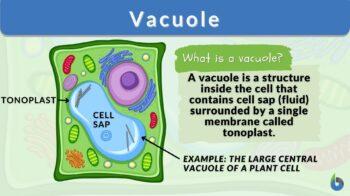
A vacuole is a single membrane-bound organelle with no definite shape or size found in both prokaryotic and eukaryotic cells. While the main function is storage, there are a variety of other roles that it serves like homeostasis, osmoregulation, cell structure maintenance, autophagy, and maintenance of pH.
By the end of this article, you should be able to not just define vacuole, but also know answers to the frequently asked questions like where is the vacuole located, do animal cells have vacuoles, what is the vacuole function in animal cells, how contractile vacuoles differ from food vacuoles, what is the permanent vacuole function in the eukaryotic cells, if all animal cell parts have vacuoles, and so on.
Its presence has been noted not just in the animal cell but also in many different cells like plant cells, bacterial cells, fungal cells, and protist cells. So, let’s begin the article and enlighten you on today’s topic of Biology Online!
Vacuole Definition in Biology
What is a vacuole? In biology, the definition of ‘vacuole‘ is a single membrane-bound organelle with no definite shape or size. It is one of the largest organelles in the cell, specifically in plant cells. It is present in different types of cells like animal cells, plant cells, fungal cells, protist cells, and bacterial cells. The structure of a vacuole is basically dependent on the individual cell requirements.
Vacuoles are associated with a range of roles in the cellular system. Some of the noted roles are intracellular secretion, excretion, storage, and digestion. The main constituent of the vacuolar system is water as vacuoles are mainly completely filled with water. They form quite a lot of the cell’s volume.
There are many organic and inorganic molecules that along with water become a part of the vacuolar solution. Because of the various roles that vacuoles serve, they are home to not just solutions but also waste products, extra water, small molecules, extra supplies, proteins, etc.
Now, that we know the definition of vacuoles, we can move ahead and learn more about their etymology, discovery, structure, types, and functions.
Etymology of Vacuole
The word vacuole is a 19th-century English word that originated from the Latin word “vacuus” meaning empty.
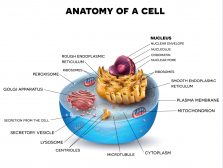
The cell and organelles
A cell is the most basic structural, functional, and biological unit of life on Earth. While we can witness some cells exist in a full-fledged manner as unicellular organisms, others form complex units and manifest the multi-cellular form of life. What constitutes and lies inside the cell hence becomes important to study and know about.
A cell is made up of many different cell organelles. These cell organelles are mostly membrane-bound structures. They tend to be associated with some specific roles and responsibilities to sustain the biological functioning of the cell.
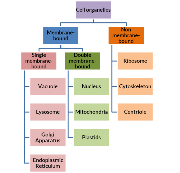
Some of the very important cell organelles found typically in eukaryotic cells are mitochondria, nucleus, Golgi apparatus, ribosomes, vacuoles, endoplasmic reticulum, vesicles, lysosomes, and cytoskeleton. Plastids and cell walls are important organelles as well but not present in animal cells.
We can divide these organelles into three different categories based on the absence, presence, and if presence then the number of cytoplasmic membrane layers the organelle is bound by. When bound by a plasma membrane, cell organelles can be single membrane-bound and double membrane-bound.
Discovery of Vacuole
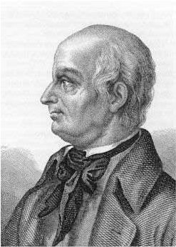
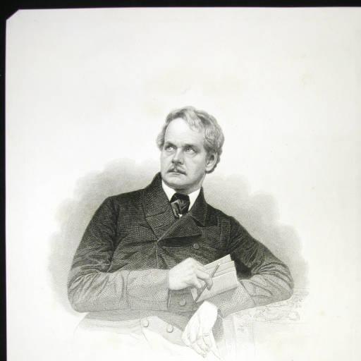
- First observation: Among the different types of vacuoles, contractile vacuoles (also called ‘stars’) were the ones to be observed for the first time. They were observed and noted by Spallanzani in 1776 in protozoan cells.
- Term coined by: The term vacuole was coined by Dujardin in 1841.
- Usage in plant biology/botany: The term vacuole was first used in Botany by Schleiden in 1842. It was used to differentiate the characteristic nature of “cell sap” from that of the “protoplasm”.
- Term ‘tonoplast’: The term tonoplast used for vacuolar membrane was coined by de Vries in 1885. The tonoplast membrane aids the maintenance of the structure of the vacuole and prevents rupture and leakage of vacuolar content.
|
|
|
|
| Figure 3: Lazzaro Spallanzani is credited with the discovery of vacuoles. He was the first person to ever observe this cell organelle. He actually observed a subtype called a contractile vacuole (Image Credit: Marianna Karamanou). Félix Dujardin, a French cytologist coined the term ‘vacuole’ (Image Credit: Archives de Parasitologie, Volume 4, 1901). Matthias Jakob Schleiden applied the term ‘vacuole’ in Botany for the first time. He is the same scientist who along with Schwann gave the “cell theory” (Image Credit: Small Portrait Collections). | ||

Watch this vid of a live Paramecium vacuole:
Biology definition:
A vacuole is a membrane-bound structure in the cytoplasm of a cell that’s primarily involved in various biological processes, such as intracellular secretion, excretion, storage, and digestion.
It is surrounded by a single membrane and contains various substances. Vacuoles for osmoregulation, for instance, contain water, ions, and other molecules. In plant cells wherein vacuoles are relatively large, the vacuole maintains internal hydrostatic pressure inside the cell and thereby helps plants by providing support to plant structures such as leaves and flowers.
As a protective container, it can hold and isolate waste products and other harmful materials from other cellular components. The vacuole also serves as storage. For example, vacuoles of seed store proteins essential for seed germination.
Etymology: from Latin vacuolum, diminutive form of vacuum
Related term: contractile vacuole
Related form: vacuolar (adjective, of, or relating to, a vacuole)
Vacuole Types
There are various types of vacuoles (membrane-bound sacs) in the biological world. Let’s look at them one at a time.
-
Gas vacuoles
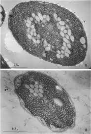
Gas vacuoles are cell organelles that are freely permeable to atmospheric gases. Their presence has been noted in many species of Cyanobacteria and only in some other bacteria and archaea. The main role associated with gas vacuoles is allowing and controlling buoyancy.

Figure 4: Gas vacuoles marked as (gv). Image Credit: Cohen-Bazire, G, 1969. -
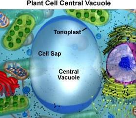
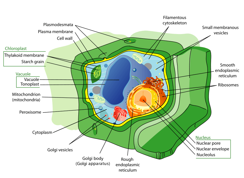
Central vacuoles
Central vacuole is terminology for the plant cell vacuoles as they have a large central vacuole occupying the majority of the cell’s volume. Cells exploit the proton gradient across the tonoplast of these central vacuoles in plant cells to transport essential nutrients into or out of the central vacuole.
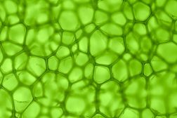
Figure 5: Note the size of the central vacuole in the plant cell. The main roles associated with central vacuoles are:
- Storage
- Maintenance of turgor pressure
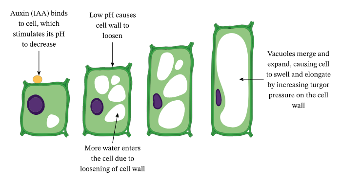
- Keeping the chloroplasts of the plant cells “closer to the cell membrane” and thereby “closer to light”: This is ensured as the central vacuole swells up and pushes all the cellular content against the cellular membrane. See Figure 6.
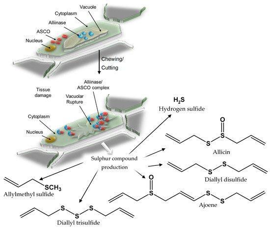
- Precursors for herbivory: Plant vacuoles store a range of chemicals in their central vacuoles that are reactive on exposure to some cytosolic content. So, when a herbivore invades the plant and tries to consume it, the vacuoles are abruptly damaged and burst open. As soon as the vacuolar chemicals react with the cytosolic content, an offensive toxin against herbivory is released. Example: Garlic has alliin and alliinase (an enzyme) separated in vacuole sap and cytoplasm. See Figure 7.

Figure 6: The central vacuole ensures that it pushes the contents of the cytoplasm including the chloroplast against the cell membrane to maximize the exposure to light. Image Credit: Kelvinsong. Figure 7: A representative flowchart of how alliin and alliinase interaction leads to the S-compounds production. The vacuolar rupture and the formation of the alliinase/ASCO complex are depicted. Image Credit: Rose, P., 2019. -
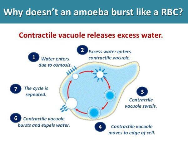
Contractile vacuoles
Contractile vacuoles are the specialized vacuoles of protist cells. They are associated with the function of osmoregulation. They carry out the removal of excessive water from the protist’s body as they tend to take in a lot of water. This is carried out by the process of contraction and expulsion.
There are 2 stages of this process. They are:
- When the contractile vacuole of protists takes in water, the vacuole swells up and increases in size. This is called the diastole stage.
- When the contractile vacuole of protists reaches saturation/threshold, the vacuole starts contracting and expelling the water out in pulses. This is called the systole stage.
Figure 8: How contractile vacuoles prevention of bursting in protists like Amoeba. Image Credit: Amoeba Sisters -
Food vacuoles
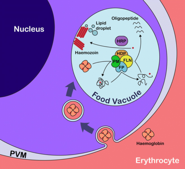
Food vacuoles are a type of vacuole that houses a number of digestive enzymes. They are therefore also referred to as digestive vacuoles. Their presence has been noted in ciliates and sporozoans. The malarial parasite, Plasmodium falciparum, also has these food vacuoles. They almost mimic lysosomes in their functioning. These vacuoles are the sites where the parasite digests the hemoglobin (Hb) of the erythrocytes. When anti-malarial drug development started, this was widely targeted and became the favorite target for scientists for such drugs.

Figure 9: The diagram shows the food vacuole carrying out hemoglobin degradation accompanied by hemozoin synthesis in the malarial parasite- Plasmodium falciparum. Image Credit: Malaria Journal.

Vacuole Structure and Functions
Let’s discuss the structure (what does the vacuole look like) and functions (roles and responsibilities) of the vacuole under separate sections.
General Structure of Vacuole
The size and shape of vacuoles vary according to the specific requirements of the cell. So, if unsure what a vacuole looks like, well, vacuoles basically have no definite shape. The vacuole is most like an amoeba in the sense that they have no fixed shape. Even the size of the vacuole isn’t fixed.
There can be one large vacuole in a cell or many small vacuoles. Plant cells usually have central vacuoles whereas animal cells typically have more than one vacuole. The structure of an animal cell and animal cell organelles is quite different from that of plant cells.
How vacuoles are formed in the cell? This question is generally common and many hypotheses have been proposed as explanations for the subject. Some scientists believe that vacuoles have formed after the fusion of many small vesicles.
Essentially, the structure of a vacuole is composed of 2 parts:
Functions of Vacuole
The vacuoles serve various different functions. Let’s learn about some of the most important ones here.
- Specialized compartment: Vacuoles serve as a specialized compartment where the cell dumps all of its extra supplies.
- Storage of toxins: Vacuoles serve an amazing role in storing and hence separating all the waste products of the cell. This separation of the toxins or toxic ions ensures that the cell and other cell organelles aren’t harmed by them.
- Maintenance of hydrostatic pressure: Vacuoles are more prominently present and identified in the eukaryotic plant cell, fungal cell, and some protist cells. The main function of the vacuole is the maintenance of an internal hydrostatic/turgor pressure inside the mature plant cells. This eventually helps in the provision of support and stature to plant structures like stems, leaves, and flowers due to the hydrostatic pressure of the central vacuole. So, this essentially answers the question, what does the vacuole do in plant cells?
-

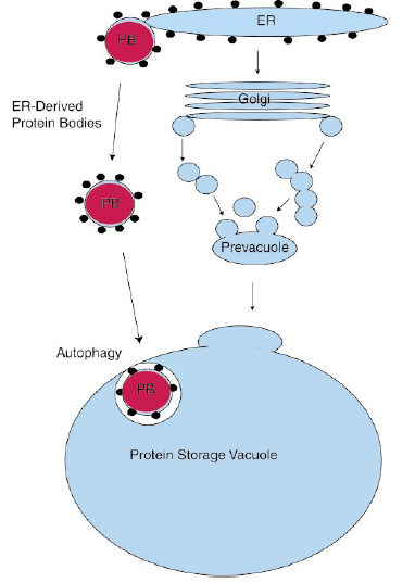
Figure 10: Protein bodies containing protein vacuoles. Image Credit: Eliot M. Hermana. Storage: Vacuole in plant cells also plays the role of storage vesicles, specifically in plant seeds. They store protein molecules in protein bodies. These proteins are essential for seed germination and hence the continuation of plant lives. Vacuole stores material within the cell; some essential (nutrients-carbohydrates, lipids, and proteins) and some non-essential (waste products, toxins) ones. It houses several digestive enzymes, small molecules, waste products, ions, unwanted substances, pigments like anthocyanin, etc. When required by the cell, vacuole releases and transports materials within the cell. Example: Vacuoles of fat cells/adipocytes in animals contain lipid molecules. See Figure 10.
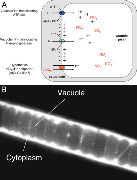
- Maintenance of pH: Vacuoles aid the optimal pH maintenance in the cellular system, i.e. in the cytoplasm. When the pH in the cell’s environment drops below the optimum level with a haphazard change in the chemical gradient, protons (or H+ ions) start moving from the external environment into the cell. This raises the acidity of the cell and holds serious implications for the cell system. So, here comes the vacuole to the cell’s rescue. The tonoplast or the vacuolar membrane helps in the facilitation of moving the protons or H+ ions from the cytoplasm to the vacuole against the concentration gradient. Although this increases the acidity of vacuole but simultaneously also ensures that the cell’s pH is maintained. See Figure 11.
- Growth: Central vacuole functions in plant cells in a very unique way. They have the ability to increase immensely in their size and thus facilitate the quick growth of plant parts by using only water. Therefore, vacuole function in a plant cell is often stated as extraordinary as growth in animal cells isn’t due to this same phenomenon. See Figure 12.
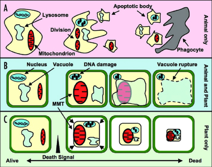
- Autophagy: Vacuoles also play an indispensable role in autophagy. They maintain a healthy balance between the two biologically contrasting processes: biogenesis and degradation. This pertains to many different substances, cell structures, misfolded proteins, invading bacteria, etc. They are known to store the by-products of the autophagy process (age-related or damage-related). See Figure 13.
- Growth site for some bacterial species: Some species of Salmonella are able to resist the acidic nature of vacuoles and also reproduce in many mammalian animals’ vacuoles after getting engulfed.
Structure and Function of Vacuoles in Animal Cells
Animal cells usually have many, small vacuoles rather than one large vacuole. An important point to note here is that not all animal cells have vacuoles, unlike all plant cells. Animal cell vacuole functions by mostly providing assistance in the processes of endocytosis and exocytosis.
The materials that enter the cell often land up in the vacuoles of the animal cells after endocytosis. Also, vacuoles are the storehouses for various protein and lipid molecules that are on the cell’s command released from vacuoles and then transported out of the cell to the extracellular environment via exocytosis.
Since animal cells have dedicated organelles called ‘lysosomes’ for the breakdown of substances, vacuoles aren’t needed for this purpose in animal cells, unlike plant cells.
Some animal cells house symbiotic bacteria in their vacuoles but this happens only in cells of some specific organs.
Structure and Function of Vacuoles in Plant and Fungi Cells
-
Plant cells
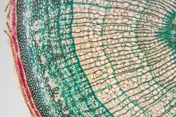
Plant cells tend to possess a large central vacuole. Plant cell with central vacuole usually takes up a large volume of the cell (commonly 30-55%, sometimes even 90-95% of cell volume). They provide support to the cell and eventually to the plant part that the cells constitute. Additionally, plant vacuoles also house a variety of molecules like secondary metabolites, pigment molecules, etc. The central vacuole of plant cells also houses a number of degradative enzymes. Another interesting point to note is that the constitution of plant cells changes during the seasons as plants can’t move (i.e. they are stationary). In the same line of thought, scientists have noted many plant cells change the organization of their vacuoles across the seasons. For example, the vascular cambium cells tend to possess a large number of small vacuoles in the winter season while all of them fuse to form one large central vacuole as the summer season arrives.
-
Fungal cells
The vacuolar structure of fungal cells is usually more similar to that of animal cells. But the functions that vacuoles of fungal cells perform are similar to that of plant cells as both fungal cells and plant cells lack lysosomes. Some of the essential functions performed by the vacuoles of the yeast cells (a type of fungi) are:
- Homeostasis of cell pH
- Enrichment and concentration of ions
- Osmoregulation
- Storage of amino acids and polyphosphates
- Degradation processes.
- Storage of toxic ions like strontium (Sr2+), lead(II) (Pb2+)
Structure and Function of Vacuoles in Bacteria Cells
An important point to note as well is that vacuoles aren’t present in all bacterial cell types. Wherever they are present, their main function is storage as in animal cells.
Example:
- Some species of sulfur bacteria possess very large-sized vacuoles (taking up to 98% of the cell’s volume).
- Some species of cyanobacteria possess a special type of vacuole that is “permeable to atmospheric gases”.
Structure and Function of Vacuoles in Protist Cells
The protist cell vacuoles are known to specifically store water. There is special terminology for the vacuoles of protists, i.e., contractile vacuoles. These were the first vacuoles of all kinds to be observed by scientists.
The contractile vacuoles help in the regulation of the amount of water in a protist cell. Hence, protist vacuoles are identified as “osmoregulators”. Since the protists living in water tend to intake excessive amounts of water, their cellular structure is prone to rupture and bursting.
In such a scenario, vacuoles come to the rescue and prevent the cell from bursting by contraction and expulsion of excess water from the cell. There can be one too many contractile vacuoles per protist cell. Also, the nature of vacuole motility varies from protist genera to genera.
Example:
- Euglena: Vacuole=stationary inside the cell.
- Amoeba: Vacuole=moves inside the cell.
Apart from this, in some protists, vacuoles serve the basic roles of digesting food and storing waste produced by the cell.
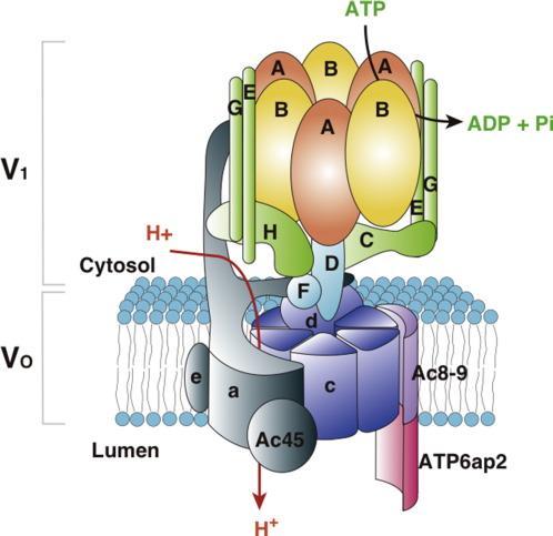
The curious case of V-ATPases in Vacuoles!
ATPases are a set of enzymes that catalyze the 2 life-sustaining, essential, reversible reactions of ATP disintegration and ATP generation.
ATP disintegration: ATP🡪 ADP + free phosphate ion (Pi)
ATP generation: ADP + free phosphate ion (Pi)🡪 ATP
There are many different types of ATPases in the biological world.

One of them is “V-ATPase” or the “vacuolar-ATPase”. This ATPase is present in the tonoplast (vacuolar membrane). It is functional in almost all types of eukaryotic cells.
While the F-ATPase that we commonly know from mitochondria and chloroplast perform the remarkable function of ATP generation at the expense of the proton-motive force, V-ATPases function exclusively as the ATP disintegration-dependent or ATP-dependent proton pumps. In V-ATPases, the energy from ATP’s disintegration drives the protons from the cytoplasm to the vacuole.
Now, you must be wondering why energy needs to be expensed for moving the protons here??
The answer to this question is in the chemical gradient of protons or H+ ions inside a cell.
The cytoplasm is usually less acidic than the vacuole. This means that the cytoplasm usually contains fewer H+ ions than the vacuole. When the protons need to be moved from a lower concentration to a higher concentration, it’s against the ‘concentration gradient’ H+ ions. Such a transfer isn’t possible spontaneously. Hence, V-ATPases by their intrinsic nature help in facilitating this process. The energy requirements for this transfer are met by the disintegration of ATP by V-ATPases.
This ability of V-ATPases in the vacuolar membrane is used to transfer protons (H+ ions) from cytoplasm to vacuole and carry out some other secondary transport processes too.
Answer the quiz below to check what you have learned so far about vacuoles.

References
- Schulz-Vogt, H. N. (2006). Vacuoles. Microbiology Monographs.
- Nelson, N., Perzov, N., Cohen, A., Hagai, K., Padler, V., & Nelson, H. (2000). The cellular biology of proton-motive force generation by V-ATPases. The Journal of experimental biology, 203(Pt 1), 89–95. https://doi.org/10.1242/jeb.203.1.89
- Matile P (1993). “Chapter 18: Vacuoles, discovery of lysosomal origin”. Discoveries in Plant Biology. Vol. 1. World Scientific Publishing Co Pte Ltd.
- Banerjee, R.; Liu, J.; Beatty, W.; Pelosof, L.; Klemba, M.; Goldberg, D. E. (2002). “Four plasmepsins are active in the Plasmodium falciparum food vacuole, including a protease with an active-site histidine”. Proceedings of the National Academy of Sciences. 99 (2): 990–995. Bibcode:2002PNAS…99..990B. doi:10.1073/pnas.022630099. PMC 117418. PMID 11782538.
- Pappas, George D.; Brandt, Philip W. (1958). “The Fine Structure of the Contractile Vacuole in Ameba”. The Journal of Biophysical and Biochemical Cytology. 4 (4): 485–488. doi:10.1083/jcb.4.4.485. ISSN 0095-9901
- Roberto Docampo, Veronica Jimenez, Noelia Lander, Zhu-Hong Li, Sayantanee Niyogi. (2013). Chapter Two – New Insights into Roles of Acidocalcisomes and Contractile Vacuole Complex in Osmoregulation in Protists, Editor(s): Kwang W. Jeon, International Review of Cell and Molecular Biology, Academic Press, 305: 69-113. ISSN 1937-6448, ISBN 9780124076952. https://doi.org/10.1016/B978-0-12-407695-2.00002-0.
- Cohen-Bazire, G., Kunisawa, R., & Pfennig, N. (1969). Comparative study of the structure of gas vacuoles. Journal of bacteriology, 100(2), 1049–1061. https://doi.org/10.1128/jb.100.2.1049-1061.1969
- Rose, P., Moore, P. K., Whiteman, M., & Zhu, Y. Z. (2019). An Appraisal of Developments in Allium Sulfur Chemistry: Expanding the Pharmacopeia of Garlic. Molecules (Basel, Switzerland), 24(21), 4006. https://doi.org/10.3390/molecules24214006
- Bucher, M., & Fabiańska, I. (2016). Long-Sought Vacuolar Phosphate Transporters Identified. Trends in plant science, 21(6), 463–466. https://doi.org/10.1016/j.tplants.2016.04.011
©BiologyOnline.com. Content provided and moderated by Biology Online Editors.