Chromatin
n., plural: chromatins
[ˈkɹəʊmətɪn]
Definition: A complex of nucleic acids (e.g. DNA or RNA) and proteins (e.g. histones)
Table of Contents
Chromatin Definition
What is chromatin in a cell? Chromatin is a complex of nucleic acids (e.g. DNA or RNA) and proteins (e.g. histones). It was identified in 1882. Initially, it was thought of as just a colored substance in a nucleus, however, later it was found that chromatin is characterized as proteins attached to DNA, and DNA was identified as the carrier of genetic information (Ma, Kanakousaki, and Buttitta 2015). Thus, we can define chromatin as a substance consisting of DNA and associated proteins (known as histones).
What are histones?
Histones are positively-charged basic proteins that bind to negatively-charged phosphate molecules of DNA. There are two major elements in a chromatin. They are the cell’s DNA and the associated proteins. The associated proteins are known as histones. Or we can say that chromatin contains proteins called histones.
Chromatin is a packaging material for DNA. If not packed well, it can tangle around itself or get damaged during cell division. The size of cells is in micrometers and a DNA can be as long as 3 meters. In order to fit such a lengthy structure into a micrometer cell, tight packing is required.
What is a nucleosome?
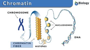
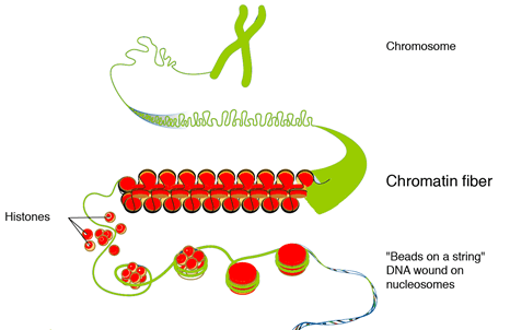
A DNA molecule wraps around the histone proteins to create tight loops known as nucleosomes.
The nucleosomes coil and are bundled together to form a kind of fiber known as chromatin fiber. These chromatins, in turn, also loop and fold around with the help of proteins to produce a chromosome. That’s why a chromosome is known to carry a part or all of the genetic material of an organism. Once the DNA is condensed into a chromosome it is now protected due to its tightly wounded structure. More details about its structure are in this section: Chromatin Structure.
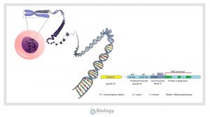
Chromatin also plays a vital role while regulating the transfer of genetic information. Before technically defining chromatin, let’s understand what it is. Let’s look at the following diagram to understand chromatins:
Chromatin is a substance made up of DNA or RNA and proteins, such as histones. It condenses during cell division (mitosis or meiosis) and becomes a chromosome. Chromatins are “unwound” condensed structures whereas chromosomes are highly packaged and more condensed than chromatins. Thus, chromosomes are the prominent form of chromatins (or genetic material) during cell division. Etymology: Greek khrōma, khrōmat- (“color”) + -in.
Chromosomes may be homologous or nonhomologous. Know more here: Difference Between Homologous Chromosomes and Sister Chromatids. Join our community and get in touch with an expert.
Where is chromatin found?
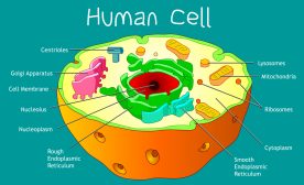
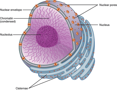
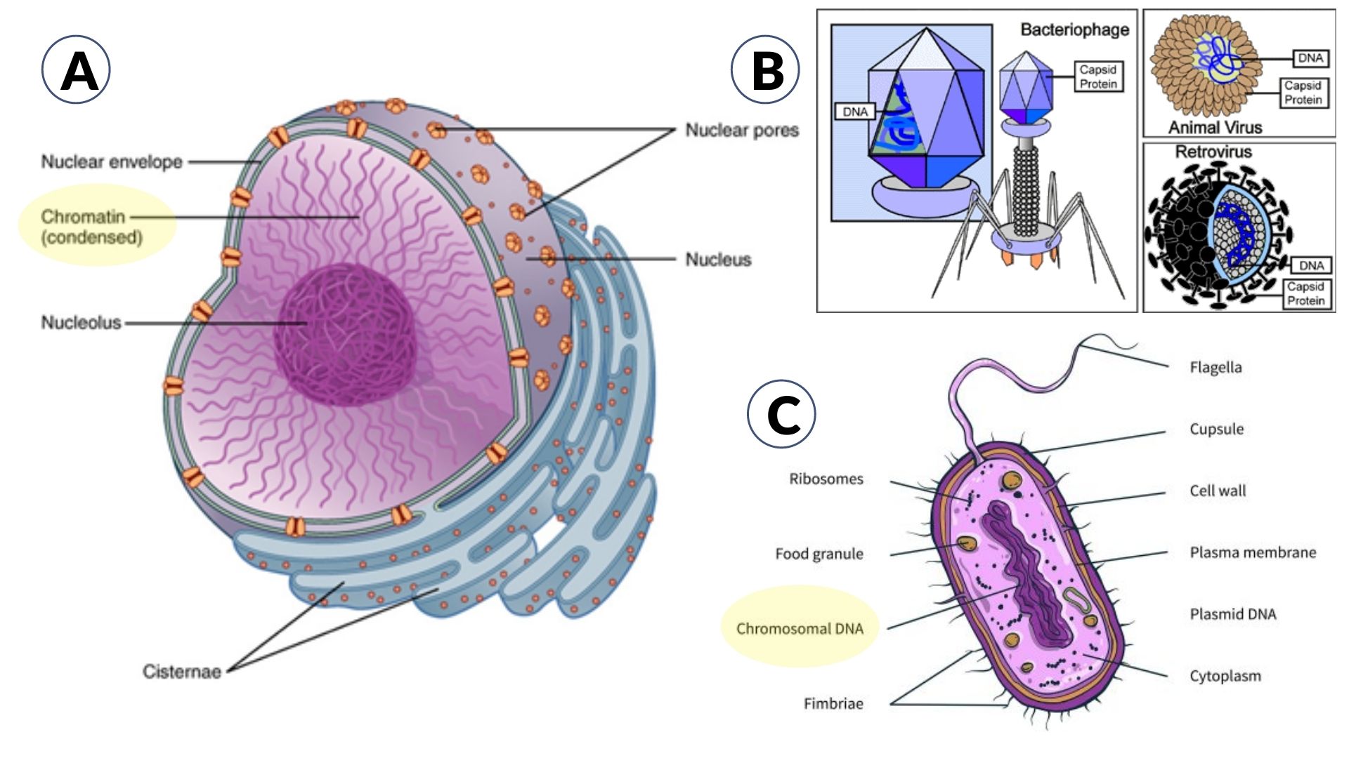
Where is the chromatin located? In eukaryotic cells, chromatin is found within the nucleus. Here is an illustration that will help you understand its location within the cell nucleus.
Genes in chromatin
The genes present in chromatin can either be turned off or on. It means in some cells a certain part of the gene is active (“switched on”) while the other is not (“switched off”). What is regulating this complex information from genes to proteins and mRNA? Yes, it’s the chromatin.
To validate this, the researchers used fruit fly as a model organism to study the on and off state of the genes in chromatin. The result of their study identified 5 distinct types of chromatin described by the unique presence of proteins.
These five types were then named as colors: Green, Yellow, Black, Blue, and Red. Black was totally inactive, green and blue were partially active, and yellow and red were fully active genes in the chromatin. They found out that the genes in Yellow chromatin were switched on in almost all the cells because they were regulating the vital functions of the cells. Red chromatins were switched on in some specific cells because they were governing more specific functions (Serra et al., 2017).
Chromatin Structure
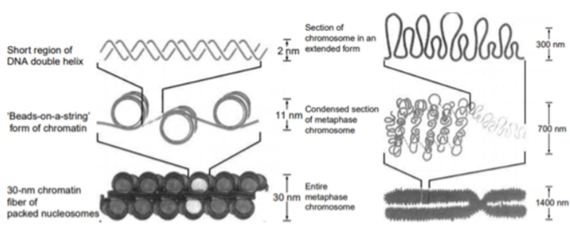
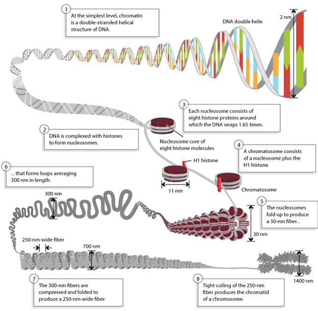
The histone protein and DNA have an equal mass in eukaryotic chromatin (although there are also some cells with non-histone proteins instead). The nucleosome is the structural unit of chromatin, which in turn, consists of DNA and (histone or non-histone) proteins. This structure is repeated throughout the genetic material of an organism. The structure of chromatin packing into the higher-order structure is shown below.
What is meant by the “beads on a string” model of chromatin?
The DNA and histone proteins provide the first level of compaction for DNA inside the nucleus. The basic unit of structure of chromatin is the nucleosome. A nucleosome is formed when DNA is wrapped around histones (the protein core) to form a “bead-like” structure. This bead-like structure is known as a nucleosome. In figure 3, the second from the top is the “beads-on-a-string” form of chromatin. The nucleosome is a complex of 146 base pairs of DNA and is wound from outside of 8 proteins, i.e. histones. Thus, DNA wrapped around histones forms a nucleosome.
There are five different types of histones, namely H1, H2A, H2B, H3, and H4. A histone core is produced when two H2A and H2B combine with H3 and H4 proteins. Approximately 145 base pairs of DNA are wrapped twice around this protein structure to form a nucleosome. The length of linker DNA can vary depending upon the species‘ gene activity and can range between 10 to 95 base pairs. There is a nucleosome after every 200 base pairs and its length was 10 nm.
When looked through a microscope, the chromatin looks like beads fitted in the string. These beads are known as nucleosomes. The nucleosome itself is composed of eight proteins known as histones. The nucleosomes form a solenoid by wrapping themselves into a 30nm spiral. In this solenoid, additional histone proteins help form chromatin structures. The chromatin condenses into chromosomes due to increasing compact structure (Baldi, Korber, and Becker 2020).
What is the relationship between DNA and chromatin?
Chromatin is the packaging of DNA. DNA and associated proteins are packed inside chromatin to fit inside a nucleus.
How DNA is assembled in chromatin structure?
There are several steps involved in the assembly of DNA into the chromatin. In the first step, H3e and H4 proteins deposit on DNA followed by H2A and H2B. A sub-nucleosomal particle is formed consisting of 146 base pairs of DNA. The second step is maturation in which ATP establishes a consistent spacing of nucleosome cores. In the next step folding of linker histones is started in a nucleofilament of 30 nm structure. In the last step further folding occurs leading to a higher level of packing. The packing ratio is about 7000.
Euchromatin vs. Heterochromatin
There are two forms of chromatin: (1) euchromatin and (2) heterochromatin. Euchromatin is less condensed and can be transcribed whereas heterochromatin is highly condensed and cannot typically be transcribed. The heterochromatin is further classified as constitutive heterochromatin and facultative heterochromatin.
The constitutive heterochromatin is the DNA sequence existing in all cells of an organism. The constitutive heterochromatin is related to the highly repeated DNA.
Similarly, facultative heterochromatin is not present in all cells. For instance, the gene encoding beta-globin in animals is present in certain cells but not in blood cells. As explained earlier, chromatin is a complex of proteins and DNA in eukaryotic cells. The nuclear DNA does not exist as linear strands but is tightly condensed and wrapped around nuclear proteins so that it can fit into the nucleus.
Chromatin Function
Initially, chromatin was considered as the substance that gives color to the cell nucleus. Later on, it was found that it is not just a coloring substance but is one of the most important DNA expression regulators. Chromatin structure has also an important role in the replication of DNA. The packaging of DNA in chromatin and nucleosome results in a tightly closed structure that is not accessible by enzymes that are responsible for the transcription, replication, and repair of DNA.
READ: How cell fixes DNA damage
The packaging of DNA structure is transcriptionally repressive and allows a basal level of gene expression only. For nucleosome structures that are open or disrupted, the DNA can more easily be replicated and transcribed.
During the transcription process, the chromatin structure is changed by some repressors and activators which interact with RNA to regulate the gene activity. Activators change the nucleosome structure resulting in RNA polymerase assembly stimulation. During replication, a similar regulation of chromatin structure occurs, which allows the replication mechanism to be in place at the origin of replication.
Chromatin also has a role in the regulation of gene expression. Using the process of position effect variegation, the genes can turn into being transcriptionally inactive by locating them near silent heterochromatic chromatins. The distance between silent heterochromatin chromatins and genes can be up to 1000 kilobase pairs. This phenomenon is referred to as epigenetic because it produces variation in phenotype.
Scientists proposed that the highly condensed nature of heterochromatin prevents the transcription of DNA. However, it is yet not fully understood how the neighboring non-heterochromatic regions are affected. The researchers found that proteins in chromatin can spread to neighboring regions to produce a similar repressive effect. The researchers also proposed that there can be some compartments in the nucleus which are not accessible to transcription factors in which heterochromatin might be residing. Thus, the chromatin in the nucleus may not be directly accessible to transcription factors.
The structure of chromatin affects DNA replication. For example, the euchromatin and other active areas of the genome replicate earlier. Similarly, in the heterochromatin and silent area around it, the replication process is also slow. Other important functions of chromatin are described below.
DNA packaging
The most important function of chromatin is the packing of long DNA strands into a much smaller space. The linear length of DNA is very long as compared to where it is residing. In order to fit safely and securely without tangling or damage, DNA needs to be compacted by some method. The compaction of DNA into the nucleus is termed condensing. The degree to which DNA is condensed inside a body is called the packing ratio. The packing ratio of DNA is about 7000. For this high level of compaction, DNA is not packaged directly into the structure of chromatin. Rather, there are several hierarchies of organization.
The initial packing is achieved by wrapping DNA around the nucleosome. This gives a packing ratio of 6. This packing is the same for both heterochromatin and euchromatin. The second level of compaction is achieved by wrapping the beads in a 30 nm fiber which is also found in both mitotic chromosomes and interphase chromatin. This wrapping increases the packing ratio from 6 to 40. The third stage of compaction is achieved by further wounding the fiber into loops, domains, and scaffolds. This final packing increased the packing ratio to 10,000 in mitotic chromosomes and 1000 in interphase chromatins.
Chromosomes are most compressed during metaphase. During the cell division of eukaryotic cells, the DNA needs to be equally divided into two daughter cells. During this phase, the DNA is highly compacted and once the cell completes the division, the chromosome uncoils again. When the length of metaphase chromosomes is compared with linear DNA, the packing ratio can be as high as 10,000:1. This high level of compaction is achieved by phosphorylation of histone H1.
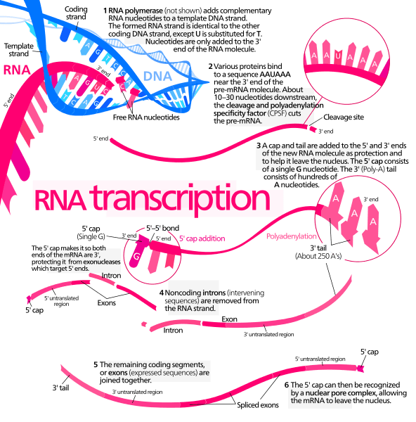
Transcription regulation
Transcription is the process of the transfer of genetic information from DNA to proteins. This information is then transcribed into RNA. The final step is the translation of RNA into functional proteins. The transcription process is controlled by chromatin. If chromatin is strengthened and restricts access to read the proteins, the transcription will stop. The heterochromatin is a condensed type of chromatin that is highly packed and proteins cannot read the DNA. While the euchromatin is not so tightly packed and proteins can conduct the process of DNA description. Similarly, there are active and inactive chromatins that can contribute to transcriptional bursting or discontinuity of transcription.
Other factors in transcription include association and dissociation of transcription factor complex present in chromatin. This phenomenon is considered the reason for the high variability in gene expression that occurs between cells in the isogenic population.
Chromatin and DNA Repair
All the DNA-based processes depend upon the packaging of DNA into the chromatin. Chromatin has the ability to change its shape and structure because of the dynamic arrangement of proteins. When DNA is damaged, chromatin relaxation occurs. This relaxation allows the proteins to bind to DNA and repair it.
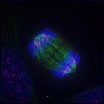
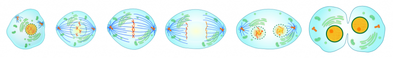
Chromatin in Mitosis
Mitosis is the process of cell division in which the resulting two cells (daughter cells) have the same type and number of chromosomes as the parent nucleus. Chromatin has an important function during the four steps of mitosis.
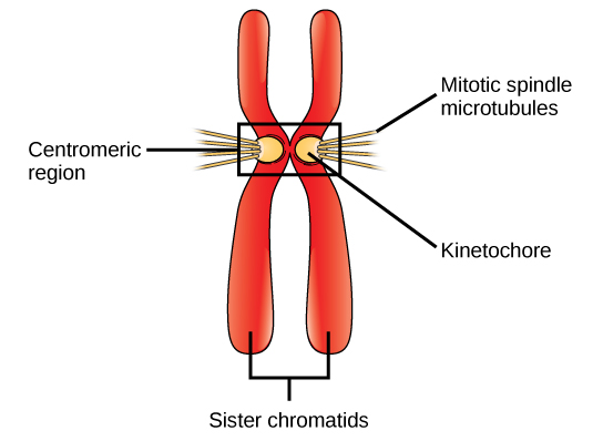
- Prophase: During this phase, the chromatin fibers wrap around to form the chromosomes. The replicated chromosome comprises two chromatids combined at the centromere.
- Metaphase: During this phase, chromatin extremely condenses
- Anaphase: During this phase, the spindle microtubules pull the two identical chromosomes to the end of the cells and separates them.
- Telophase: In this phase, the new chromosomes are separated into their own nucleus. At this point, chromatin fibers become less condensed by uncoiling. Two identical cells are produced with the same number of chromosomes.
Chromatin, Chromosome, and Chromatid
Although all the three structures i.e. chromatin, chromosomes, and chromatids are present in the nucleus of the cell and are composed of DNA, however, they are uniquely identified as described below:
Chromatin vs. Chromosome
The major difference between chromatin and chromosomes is that chromatin is composed of DNA and histones that are packed into a fiber whereas a chromosome is a single-stranded form of condensed chromatin. The chromosome structure is based on the fine fiber of chromatin. While chromatin functions are described above, the chromosomes function is vital during mutation, regeneration, cell division, variation, and heredity. Moreover, during cell division, chromatin condenses to form a chromosome and the chromosome is double-stranded with an X shape. The two strands are connected to the center through a region known as the centromere.
Where in the cell are chromosomes located?
The chromosomes are present in the nucleus of a eukaryotic cell. In prokaryotes, the chromosome is typically a single loop of stable chromosomal DNA in the nucleoid, e.g. of a bacterial cell. Prokaryotic DNA is associated with non-histone proteins. In viruses, there is also no nucleus and so the chromosome may appear as a short linear or circular structure of DNA or RNA molecule often lacking any structural proteins encased by an envelope or a capsid of its head.
What is the relationship between chromatin and chromosomes?
The relation between chromatin and chromosome is that chromatin further undergoes condensation to form a chromosome. A chromosome DNA packing ratio is higher than chromatin.
Chromatin vs. chromatid
There are two strands of chromosomes. The single strand of the chromosome is called a chromatid. These chromatids separate at the end of cell division to become daughter chromosomes. Thus chromatin is entirely different from chromatid because the major elements of chromatin are DNA and associated proteins in the form of fiber while chromatid is a part of the chromosome. Yes, the chromatid contains chromatin.
Chromatin vs. nucleosome
The nucleosome is the part of DNA that is wrapped around the core of proteins. Chromatin is the DNA complex with protein and helps in DNA condensation for packing into the nucleus.
Try to answer the quiz below to check what you have learned so far about chromatin.

References
- Anthony T. Annunziato. 2008. “DNA Packaging: Nucleosomes and Chromatin.” Nature Education. 2008. https://www.nature.com/scitable/topicpage/dna-packaging-nucleosomes-and-chromatin-310/#%0Ahttps://www.scribd.com/document/257368023/DNA-Packaging-Nucleosomes-and-Chromatin-Annunziato-2014.
- Baldi, Sandro, Philipp Korber, and Peter B Becker. 2020. “Beads on a String—Nucleosome Array Arrangements and Folding of the Chromatin Fiber.” Nature Structural & Molecular Biology 27 (2): 109–18.
- Creative-diagnostics. 2017. “The Structure and Function of Chromatin.” 2017. https://doi.org/10.7326/0003-4819-83-3-445_5.
- Jansen, A., and K. J. Verstrepen. 2011. “Nucleosome Positioning in Saccharomyces Cerevisiae.” Microbiology and Molecular Biology Reviews 75 (2): 301–20. https://doi.org/10.1128/mmbr.00046-10.
- Ma, Yiqin, Kiriaki Kanakousaki, and Laura Buttitta. 2015. “How the Cell Cycle Impacts Chromatin Architecture and Influences Cell Fate.” Frontiers in Genetics 6: 19.
- Serra, François, Davide Baù, Mike Goodstadt, David Castillo, Guillaume J Filion, and Marc A Marti-Renom. 2017. “Automatic Analysis and 3D-Modelling of Hi-C Data Using TADbit Reveals Structural Features of the Fly Chromatin Colors.” PLoS Computational Biology 13 (7): e1005665.
©BiologyOnline.com. Content provided and moderated by BiologyOnline Editors.