Anatomy
n., [əˈnætəmi]
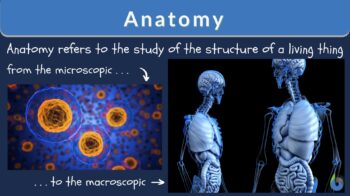
Definition: the study of the structure of the body of an organism
Table of Contents
Anatomy Definition
Often we hear the term ‘anatomy‘ while learning about the body and how it works. What is anatomy? What does anatomy mean, how would we define anatomy? Anatomy is the study of a specific biological branch in science that deals with the structure and identification of organism’s bodies and their different sections. Though the phrase “anatomy of the body” is often used in reference to humans and human body parts, it includes all living things. This study of the body structure is divided into two subsections of gross or macroscopic anatomy and microscopic anatomy.
History of Anatomy
Humans are a curious species and we have always been interested in how bodies have been formed and created. Anatomical structures, hyroglifics, and drawings that date back as early as 750000 BCE have been discovered in caves. These showed evidence that the primitive human had a relatively vast knowledge of the anatomy of the human body, despite the simplicity of the drawings of what was seen.
It can also be confirmed that even late in the Paleolithic Period, humans had begun to perform small “medical” procedures. A small hole was made into the skull of a person who may have been suffering from mental diseases. Though the reasoning behind it was not always scientific, some of the persons who underwent these procedures survived and showed a breakthrough in this branch of medicine.
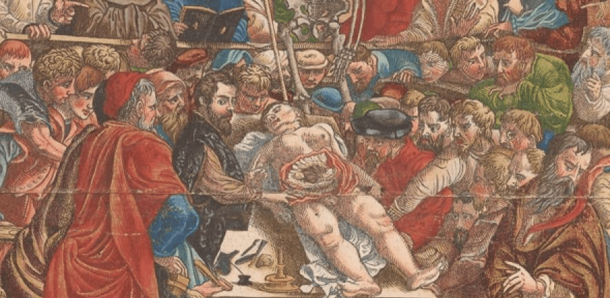
Fast forward to the Ancient Romans who used treating their injured gladiators to gain more anatomical knowledge. However, this could only be done secretly as the dissection of human bodies was not allowed. Hence, the Romans depended on using animal anatomy and bodies to further compare and contrast their findings on humans. During this period, the researcher and experimentalist Galen became a practicing physician. He studied macroscopic anatomy of which most were dissections of animals. The findings he made would carry on for centuries to come.
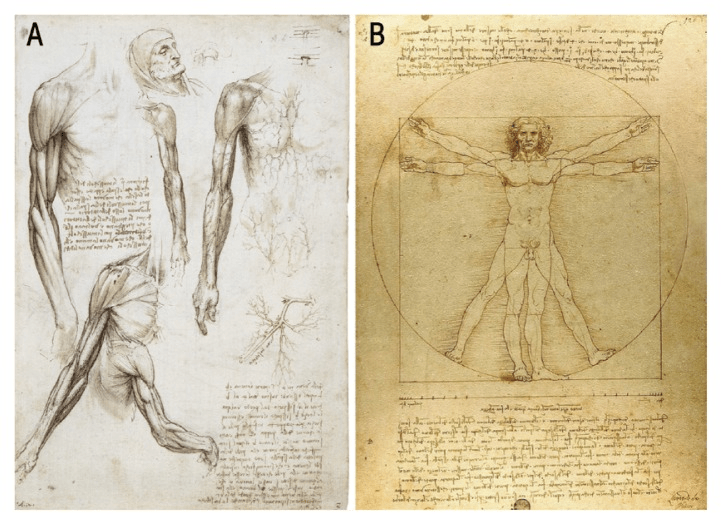
The Renaissance Period brought about the beginning of anatomy charts, sketches by great artists such as Leonardo Da Vinci and Rembrandt van Rijn. These artists brought about some of the cornerstones for the modern anatomical sketches and human body diagrams of the organ systems of the body. The artists worked in conjunction with scientists to draw anatomically accurate sketches after the dissection of parts of human bodies. This period also brought about the work of Andreas Vesalius who contributed extensively to anatomy by his constant study of dissected humans and his work on the first-ever book on human anatomy.
From the 17th to 20th centuries, numerous anatomists and physicians have contributed to creating the anatomy that we learn today and aiding in the advancement of medical knowledge. Up until those times, there was discrimination against scientists who wanted to use cadavers or dissect living humans in order to study human internal organs and human systems. Abraham Flexner wrote his famous article from the 20th century that highlighted the importance of medical research using the organs in the body and how dissections contribute to basic medical education and training. This helped to bring about normalcy in the field of anatomy and push the medical discoveries that we know today.
Important Figures in Anatomy
Fields in biology often become more diverse due to research done by numerous scientists, doctors, and medical researchers who delve more into those studies. An expert in anatomy or an anatomist may do studies in complete anatomy or specific areas. Many historical anatomists have contributed to what we call anatomy today.
William Harvey was an English physician who lived between 1578 and 1657. He is best known for being the official doctor for King James the first and discovering the circulatory system. Though before Harvey, others have hypothesized that there was a method by which blood is pumped around the body, Harvey confirmed these theories with arguments and experiments. Harvey used the volume to measure and estimate the amount of blood that would pass through the human body organs at a given timeframe. Since the body could not produce such large quantities of blood at once, it had to be circulated. Furthermore, he investigated the heartbeat and how it worked as a pump to push the blood around the body. After publishing in 1649 in a medical journal, Harvey’s work received much criticism but was eventually accepted.
Another well-known anatomist is Andreas Vesalius. He lived in Greece until the age of 50 where he studied and extensively described human anatomy and physiology. After receiving his doctorate, Vesalius devoted much of his time to dissecting and observing cadavers. So intrigued by this, Andreas began to follow a pattern of study involving doing his dissections and studying the ancient scientific texts in detail. He also discovered that the Galenic anatomy, which was used in many universities, was based on animal anatomy rather than human body systems. This was possibly due to the fact that human dissection was forbidden in the past. This discovery helped to push his creation of one of the first anatomical textbooks ‘Fabrica’ that was printed in 1543.
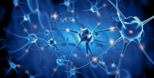
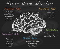
French anthropologist and pathologist, Paul Broca, studied lesions in the brain that helped modern biology understand cranial functions better. He discovered in 1861 what is now known as the convolution of Broca, a section in the left frontal lobe of the brain known for helping with the articulation of speech. This also made him the first person to prove that specific parts of the brain were related to certain functions in the systems of the human body.
Georges Cuvier was the founding father of comparative anatomy and paleontology. His work on the study of marine invertebrates as a tutor was sent to the Museum of Natural History in Paris where he was then encouraged to become a member of their team. He was the first to break down animals into subcategories based on their systems in the body. This rejected the typical anatomical organization that was used prior to the 18th century and helped others to explore the fact that animals are anatomically different and why this could be. All this and more helped Curvier to build the foundation of paleontology as a science.
Gross Anatomy
One of the subdivisions of anatomy is gross anatomy. This deals with the study of anatomy that you can see with the naked eye on a macroscopic level. Gross anatomy complements cytology and histology. Cytology involves the study of cells as the first basic unit of life whereas histology is the study of tissues and their structures. Gross anatomy and growth and development often go hand in hand since it describes the development of each trait of an organism’s body part.
Macroscopic anatomy is further divided into surface, regional and systemic anatomy. These deal with outside of the body, regions of the body and specific body systems respectively. When we study surface anatomy, we do not need to dissect to learn and observe. This subdivision looks at the external body form and what it does in order to allow the body to function while protecting the internal structures. Regional anatomy looks at specific sections of the body and how they work together to carry out numerous functions. The gastrointestinal system and the circulatory system are both examples of the eleven (11) body systems that are focused upon when dealing with systemic anatomy.
Microscopic Anatomy
The study of the structure of organisms at a microscopic level is microscopic anatomy. Microscopic anatomy is sometimes used interchangeably with histology but that is incorrect as microscopic anatomy includes both histology and cytology. Histology studies how cells are the building blocks of the body and so develop from cells to tissues into organs and organ systems. These different levels of development come together to create a living thing. However, microscopic anatomy deals with only tissues and smaller entities since these are the only ones that can fit under the microscope.

As said above, microscopic anatomy involves both histology and cytology. Both of these involve the thin slicing of organs to obtain specimens for the microscopes. This can be done on either live or dead cells and tissues. These are then dyed in order to obtain a contrast and visibility between different organelles and components of the cells and tissues. This method makes the studying of the anatomy of miniature body parts easier to do.
Other Branches of Anatomy
Aside from macroscopic and microscopic anatomy, there are many other branches of anatomy. The five main ones are embryology, developmental anatomy, radiographic anatomy, and pathological anatomy.
As the name implies, embryology is the study of the embryo of an animal. An embryo exists during the period of time from which the egg is fertilized to the eighth week of the life of an organism. It looks critically at the organism’s cell differentiation, division, and gastrulation. It is often described as the basis for the comprehension of how nervous systems and other crucial parts of the animal develop and function. This study has also in recent times, helped with the study of stem cells and how they can relate to cancer.
The study of developmental anatomy is a wider and longer study than embryology as it studies from the moment of fertilization all the way into adulthood. This study puts the anatomy meaning into perspective as it concentrates on all aspects of the body form. It is a closer look at how the organism’s body will change over the course of its life.
Radiographic anatomy uses radiology in the form of x-rays to study the body and all its systems and organs. These can be radiographs or computerized tomography (CT) scans. These take the three-dimensional form of the body and put it into a two-dimensional picture so that the real-time look of the systems can be observed by a radiologist or medical doctor.
The study of anatomy as it is altered by disease is known as pathological anatomy. This study helps the diagnosis and treatment of countless diseases through microscopic analysis of samples from bodily fluids, tissues, organs, and sometimes the entire body or autopsy.
Human Anatomy
This is the branch of anatomy that is most popular throughout the world and for obvious reasons. We as humans are always curious as to how our body works and how it functions. However, before you know how the parts of the body work you need to know the structure or form. This is where human anatomy comes in. The way the human body is organized is a special system in itself. We start from the cell, which is the building block of the body. Cells come together to form tissues, tissues form organs that combine to form organ systems. These 11 body systems then form the human body.
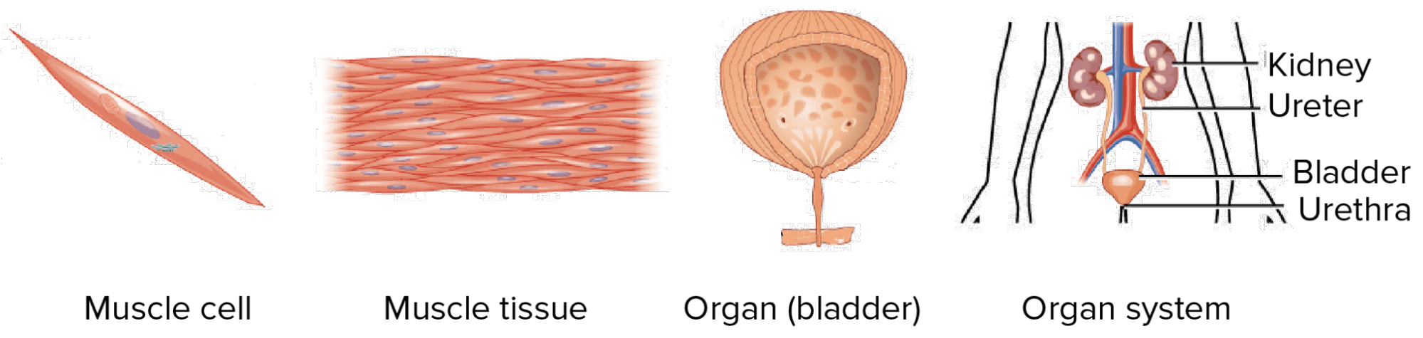
Cells are the smallest unit in the body and every other body part is made up of cells. The fundamental type of cell is the stem cell, which can develop into any kind of specialized cell needed by the body. There are over 200 types of specialized cells in the human body — ranging from reproductive cells that assist in the reproductive creation of offspring to red blood cells that carry oxygenated and deoxygenated blood throughout the body.
Groups of cells with similar functions that come together to function as a unit are known as tissues. There are 4 main types of tissues; they are epithelial, muscle, connective, and nervous. Epithelial tissues are the main tissues of the glands and have multiple functions including absorption, secretion, and filtration. Connective tissues bind structures together, creating a solid support system and protecting against diseases. The tissues that are specially designed to assist with the movement of the body are the muscle tissues. These include the cardiac tissues which contract and relax the heart, allowing blood to be pumped around the body. The brain, spinal cord, and nerves consist of nervous tissues. These are responsible for controlling the body and its functions, which includes coordination.
When several types of cells and tissues come together they form organs. Organs are the structures that perform specific functions. The lung’s purpose is to facilitate gas exchange so that the blood cells can receive oxygen and release carbon dioxide. The skin serves as a barrier for invaders, protects and regulates temperature among other things. When these organs work in conjunction with other organs they form organ systems.
There are a total of eleven (11) organ systems. These systems include the following:
- cardiovascular system (circulatory system)
- digestive system (GI tract)
- endocrine system
- integumentary system
- lymphatic system
- muscular system
- nervous system
- skeletal system
- reproductive system
- respiratory system
- urinary system
Each of these contributes in its own way to the anatomy and physiology of the body. The immune system is another biological system but overlaps another system, particularly, the lymphatic system.
Learn the different functions of the human body systems here: The Human Physiology – Biology Tutorial.
Watch the video below for the human body systems overview and functions.
Anatomical Nomenclature
As the branch of anatomy continued over periods of time, a specific way of referring to certain items and areas in an organism’s body was developed. This is called anatomical nomenclature and it is used by both researchers and medical doctors alike. This nomenclature is only available officially in its Latin form. Anatomical nomenclature was created because when referring to the human body, terms like ‘up’ and ‘down’ can become confusing depending on the position of the body. Hence, this will help researchers and medical personnel to be able to follow along while someone else is describing the position of a body part.
A human standing erect, on both legs, facing front with the palms facing forward is considered to be in the standard anatomical position. Using this standard position, many things can be described using the anatomical nomenclature, such as superior meaning towards the head or even inferior meaning towards the feet.
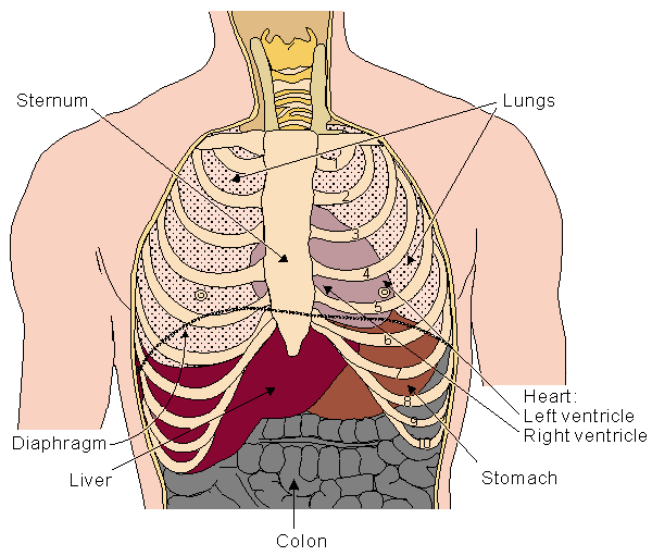
For instance, one would say that the heart is posterior to the sternum, meaning that anatomically, the heart is ‘at the back of’ the sternum. Also, saying that the hand is part of the superior extremity is saying that the hand is part of the upper part of the body (i.e., upper limb) although it is also towards the end of the upper limb.
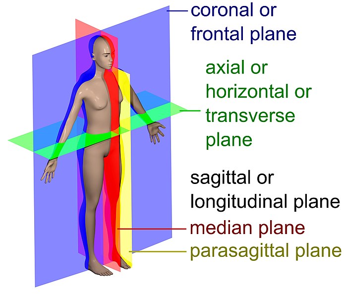
In the diagram below, the different anatomical planes are shown. An anatomical plane refers to the hypothetical plane to describe the location of a particular bodily structure. It may also be used in describing anatomic movement directions. Examples: coronal (frontal) plane, axial (horizontal or transverse) plane, sagittal (or longitudinal plane), median plane, and parasagittal plane.
Or, watch the video below.
Try to answer the quiz below to check what you have learned so far about anatomy.

References
- Anatomy Next. 2021. Understanding Human Anatomy. Anatomy Next. https://anatomy.net/blog/human-anatomy/
- Britannica, T. Editors of Encyclopaedia. 2018, November 20. Cytology. Encyclopedia Britannica. https://www.britannica.com/science/cytology
- Britannica, T. Editors of Encyclopaedia. 2020, August 19. Georges Cuvier. Encyclopedia Britannica. https://www.britannica.com/biography/Georges-Cuvier
- Britannica, T. Editors of Encyclopaedia. 2021, July 5. Paul Broca. Encyclopedia Britannica. https://www.britannica.com/biography/Paul-Broca
- Cengage. 2018. Anatomical Nomenclature. Medicine. https://www.encyclopedia.com/medicine/diseases-and-conditions/pathology/anatomical-nomenclature
- Florkin, M. 2021, May 28. Andreas Vesalius. Encyclopedia Britannica. https://www.britannica.com/biography/Andreas-Vesalius
- Gregory, A. 2021, May 30. William Harvey. Encyclopedia Britannica. https://www.britannica.com/biography/William-Harvey
- Habbal, O. 2017. The Science of Anatomy. Sultan Qaboos University Medical Journal. https://www.ncbi.nlm.nih.gov/pmc/articles/PMC5380415/
- Hefner, J. T., Linde, K. C. 2018. Atlas of Human Cranial Macroscopic Traits. Science Direct. https://www.sciencedirect.com/science/article/pii/B978012814385800001X
- Kachlík, D., Musil, V., & Baca, V. (2017). A plea for extension of the anatomical nomenclature. Part 1: Nervous system and senses. Folia morphologica, 76(2), 168-177. https://link.springer.com/article/10.1007/s00276-008-0357-y
- National Cancer Institution. 2021. Body Tissues. Anatomy and Physiology. https://training.seer.cancer.gov/anatomy/cells_tissues_membranes/tissues/
- Nature. 2021. Anatomical Nomenclature. Nature. https://www.nature.com/articles/101130a0#citeas
- Open Learn. 2020. What is Histology. Histology, microscopy, anatomy and disease. https://www.open.edu/openlearn/ocw/mod/oucontent/view.php?id=65372§ion=1
- Stoppler, M. C. 2021. Definition of Microscopic Anatomy. RxList. https://www.rxlist.com/microscopic_anatomy/definition.htm
- Teach me anatomy. 2021. Embryology. The basics of Anatomy. https://teachmeanatomy.info/the-basics/embryology/gastrulation/
©BiologyOnline.com. Content provided and moderated by Biology Online Editors.