Cell Structure
The human cell – a eukaryotic cell with many cytoplasmic structures bounded by biological membranes
Table of Contents
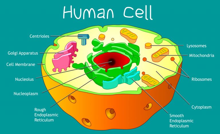
The interior of human cells is divided into the nucleus and the cytoplasm. The nucleus is a spherical or oval-shaped structure at the center of the cell. The cytoplasm is the region outside the nucleus that contains cell organelles and cytosol, or cytoplasmic solution. Intracellular fluid is collectively the cytosol and the fluid inside the organelles and the cell nucleus.
Membranes
Membranes are the gateways to the cell. The cell membrane (or plasma membrane) is the selective barrier surrounding the cell. It provides a barrier to the movement of molecules between the intra- and extracellular fluids as the cell membrane surrounds the cell, thereby, separating cellular contents from the surrounding environment.
The plasma membrane also serves to anchor adjacent cells together and to the extracellular matrix. Various signals and inputs can alter the sensitivity and permeability of membranes.
The Fluid Mosaic Model: membrane structure
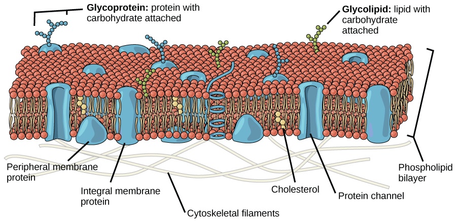
Membranes are made of a double layer of lipids, mainly phospholipids, containing embedded proteins. The embedded proteins are important as facilitators in moving molecules through the membrane. The membrane itself is organized into a bimolecular layer, meaning that the non-polar region is organized in the middle (away from water as it is hydrophobic) and the polar regions are oriented toward the outside: the extracellular fluid and the cytosol.
Another way to think of it is two rows of pins with their heads to the outside and the needle part to the inside. Heads, needles, needles, heads. Like a sandwich. As the phospholipid molecules are not chemically bound to each other and thus each molecule is free to move independently, the overall bi-layer structure has a flexible fluidity. Cholesterol molecules are also embedded in the plasma membrane and serve to deliver substances to cell organelles by forming vesicles.
The proteins embedded in the membrane are categorized into two classes:
- Peripheral membrane proteins are proteins on the membrane surface, mainly the cytosolic side where they interact with cytoskeletal elements in order to influence cell shape and motility. These proteins are not amphipathic and are bound to polar regions of the integral proteins.
- Integral membrane proteins span the entire width of the membrane, thus crossing through both the polar and non-polar regions of the structure. These proteins cannot be removed from the membrane without disrupting the lipid bilayer.
It is important to realize that membrane functions are dependent on the chemical composition and any asymmetries in composition between the two surfaces of the membrane and the specific proteins that are attached to or associated with the membrane. The plasma membrane also has an extracellular surface layer of monosaccharides that are linked to the membrane lipids and proteins. This layer is called the glycocalyx and is important in the intercellular recognition process.
Membrane Junctions
Integrins are transmembrane proteins that bind to specific proteins in the extracellular matrix and to membrane proteins in adjacent cells. Integrins help to organize cells into tissues. They are also responsible for transmitting signals from the extracellular matrix to the cell interior.
If two cells are adjacent, but separated, they may be junctured by desmosomes. Desmosomes are dense accumulations of protein at the cytoplasmic surface of the plasma membranes of both separate cells. They are infiltrated with protein fibers that extended into either cell. The purpose and function of desmosomes is to hold adjacent cells firmly in place in areas that are subject to stretching, such as skin.
Another type of membrane junction is the tight junction. These junctions are formed by the actual physical joining of the extracellular surfaces of two adjacent plasma membranes. Tight junctions are important in areas where more control over tissue processes is needed, such as the epithelial cells in the intestine that are involved in absorption.
Finally, gap junctions are actual protein channels that link the cytosols of adjacent cells. The drawback to this ‘direct link’ is that it only permits smaller molecules to pass through.
Cell Organelles
Cell organelles are the little workhouses within the cell. All the functions of life take place in each individual cell. Organelles can be released by breaking the plasma membrane, through homogenization and ultracentrifuging the mixture. The organelles are of different sizes and densities and will settle out at specific rates.
The nucleus is in the center of most cells. Some cells contain multiple nuclei, such as skeletal muscle, while some do not have any, such as red blood cells. The nucleus is the largest membrane-bound organelle. Specifically, it is responsible for storing and transmitting genetic information. The nucleus is surrounded by a selective nuclear envelope. The nuclear envelope is composed of two membranes joined at regular intervals to form circular openings called nuclear pores. The pores allow RNA molecules and proteins modulating DNA expression to move through the pores and into the cytosol. The selection process is controlled by an energy-dependent process that alters the diameter of the pores in response to signals. Inside the nucleus, DNA and proteins associate to form a network of threads called chromatin. The chromatin becomes vital at the time of cell division as it becomes tightly condensed thus forming the rodlike chromosomes with the enmeshed DNA. Inside the nucleus is a filamentous region called the nucleolus. This serves as a site where the RNA and protein components of ribosomes are assembled. The nucleolus is not membrane-bound, but rather just a region.
Ribosomes are the sites where protein molecules are synthesized from amino acids. They are composed of proteins and RNA. Some ribosomes are found bound to the granular endoplasmic reticulum, while others are free in the cytoplasm. The proteins synthesized on ribosomes bound to the granular endoplasmic reticulum are transferred from the lumen (open space inside the endoplasmic reticulum) to the Golgi apparatus for secretion outside the cell or distribution to other organelles. The proteins that are synthesized from free ribosomes are released into the cytosol.
The endoplasmic reticulum (ER) is collectively a network of membranes enclosing a singular continuous space. As mentioned earlier, granular endoplasmic reticulum is associated with ribosomes (giving the exterior surface a rough, or granular appearance). Sometimes granular endoplasmic reticulum is referred to as rough ER. The granular ER is involved in protein synthesis as well as in packaging proteins for the Golgi apparatus. The agranular, or smooth, ER lacks ribosomes and is the site of lipid synthesis. In addition, the agranular ER stores and releases calcium ions Ca2+.
The Golgi apparatus is a membranous sac that serves to modify and sort proteins into secretory/transport vesicles. The vesicles are then delivered to other cell organelles and the plasma membrane. Most cells have at least one Golgi apparatus, although some may have multiple. The apparatus is usually located near the nucleus.
Endosomes are membrane-bound tubular and vesicular structures located between the plasma membrane and the Golgi apparatus. They serve to sort and direct vesicular traffic by pinching off vesicles or fusing with them.
Mitochondria are some of the most important structures in the human body. They are the site of various chemical processes involved in the synthesis of energy packets called ATP (adenosine triphosphate). Each mitochondrion is surrounded by two membranes. The outer membrane is smooth, while the inner one is folded into tubule structures called cristae. Mitochondria are unique in that they contain small amounts of DNA containing the genes for the synthesis of some mitochondrial proteins. The DNA is inherited solely from the mother. Cells with greater activity have more mitochondria, while those that are less active have less need for energy-producing mitochondria.
Lysosomes are bound by a single membrane and contain highly acidic fluid. The fluid acts as digesting enzyme for breaking down bacteria and cell debris. They play an important role in the cells of the immune system.
Peroxisomes are also bound by a single membrane. They consume oxygen and work to drive reactions that remove hydrogen from various molecules in the form of hydrogen peroxide. They are important in maintaining the chemical balances within the cell.
The cytoskeleton is a filamentous network of proteins that are associated with the processes that maintain and change cell shape and produce cell movements. The cytoskeleton also forms tracks along which cell organelles move propelled by contractile proteins attached to their various surfaces. Like a little highway infrastructure inside the cell. Three types of filaments make up the cytoskeleton.
Microfilaments are the thinnest and most abundant of the cytoskeleton proteins. They are composed of actin, a contractile protein, and can be assembled and disassembled quickly according to the needs of the cell or organelle structure.
Intermediate filaments are slightly larger in diameter and are found most extensively in regions of cells that are going to be subjected to stress. Desmosomes in the skin will contain filaments. Once these filaments are assembled they are not capable of rapid disassembly.
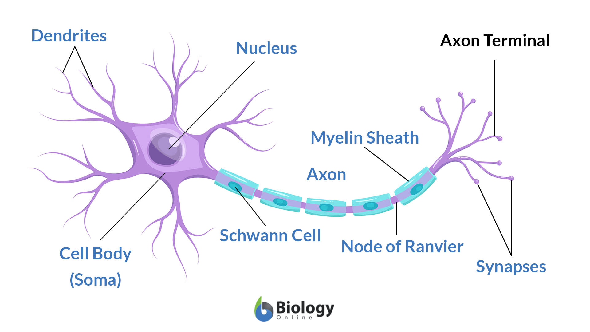
Microtubules are hollow tubes composed of a protein called tubulin. They are the thickest and most rigid of the filaments. Microtubules are present in the axons and long dendrite projections of nerve cells. They are capable of rapid assembly and disassembly according to need. Microtubules are structured around a cell region called the centrosome, which surrounds two centrioles composed of 9 sets of fused microtubules. These are important in cell division when the centrosome generates the microtubular spindle fibers necessary for chromosome separation.
Finally, cilia are hair-like motile extensions on the surface of some epithelial cells. They have a central core of 9 sets of fused microtubules. In association with a contractile protein, these microtubules produce movement in cilia. Ciliar movements propel the luminal contents of hollow organs lined with ciliated epithelium.
Now, those are the fundamental characteristics of a typical human cell. Take note though that different cell types have different characteristics. These features make these cells capable of doing specific functions, which is crucial to cell growth and cell survival.
See these figures as examples:
If you would like to know the difference between human cells (in terms of basic structure, cell biology, and major functions) from other eukaryotic cells (such as plant cells and animal cells), read these modules:
- Plant cells – a module about the cells of plants characterized by having cell walls and plastids
- Animal cells – a module about the cells of animals devoid of cell walls and plastids
- Plants vs animal cells – tutorial about the differences between plants and animal cells
- Cell theory – a module about the various theories that explain the idea of organismal constitution, cell structure, and function.

Fun activity: Animal Cell Coloring worksheet – get acquainted with the different animal cell parts.
You will also like...
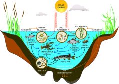
Freshwater Producers and Consumers
Freshwater ecosystem is comprised of four major constituents, namely elements and compounds, plants, consumers, and deco..
Regulation of Biological Systems
Regulation of Biological Systems tutorials are focused on the modulation of biological systems from cell to population l..

Meiosis and Alternation of Generations
Plants are characterized by having alternation of generations in their life cycles. This tutorial is a review of plant m..
Chemical Composition of the Body
The body is comprised of different elements with hydrogen, oxygen, carbon, and nitrogen as the major four. This tutorial..

Animal Growth Hormones
Hormones are produced in the endocrine glands of animals. The pituitary gland and hypothalamus are the most impor..
The Conscious & Unconscious Nervous System
This tutorial elaborates on how the nervous system works, particularly at the tissue level of the brain. There are three..